Skip to content
recaplica

    One moment: security check

    Cloudflare wants to make sure you're not a robot. Tick the box below and your search will continue on its own.

    IT
    recaplica Muscular system: how the muscles in your body work
    © 2026 Recaplica · recaplica.com — All rights reserved
    Home › Science

    Muscular system: how the muscles in your body work

    By Recaplica Newsroom · Updated on September 2, 2026

    What to print

    Page numbers appear when printing with default margins.

    Slides

    Choose a cut

    Flash10 slidesThe essential thread, to present in classFull18 slidesEvery chapter and the deeper detail

    Both come with speaker notes.

    Telegram channel
    recaplica Clear in 30 seconds, yours in 10 minutes.
    In 30 seconds Key points Figures Deep dive Slides Myths Mind map Quiz Flashcards FAQ

    In 30 seconds quick read

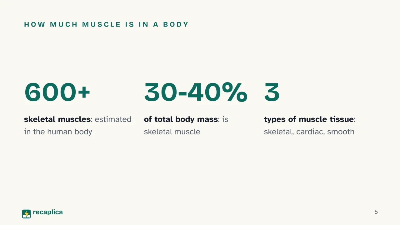
    The muscular system is the set of muscles that lets you move, breathe and keep your heart beating: the human body has more than 600 of them, and together they make up roughly 30-40% of total body mass. There are three types of muscle tissue — skeletal, cardiac and smooth — each with a different cell structure and a different degree of conscious control. Skeletal muscle is organised into progressively smaller units down to the sarcomere, where two proteins, actin and myosin, slide past each other and shorten the fibre. Contraction starts with a nerve signal, moves through a release of calcium, and runs on energy from ATP.

    Key Points

    • The human body has more than 600 skeletal muscles, which together make up about 30-40% of total body mass.
    • There are three types of muscle tissue: skeletal (voluntary), cardiac and smooth (both involuntary).
    • Skeletal muscle is organised like nested boxes: bundle, fibre, myofibril, sarcomere — the sarcomere is the unit that actually shortens.
    • Contraction starts with a nerve signal that releases acetylcholine, lets calcium into the fibre, and allows actin and myosin to slide past each other.
    • ATP is the fuel that lets myosin detach from actin and repeat the attachment cycle for as long as force is needed.
    • Only skeletal muscle moves by conscious choice: cardiac and smooth muscle work on their own, without any input from your thoughts.

    Key figures

    • 600+ the estimated number of skeletal muscles in the human body Source: Cleveland Clinic
    • 30-40% the share of total body mass made up of skeletal muscle Source: Cleveland Clinic
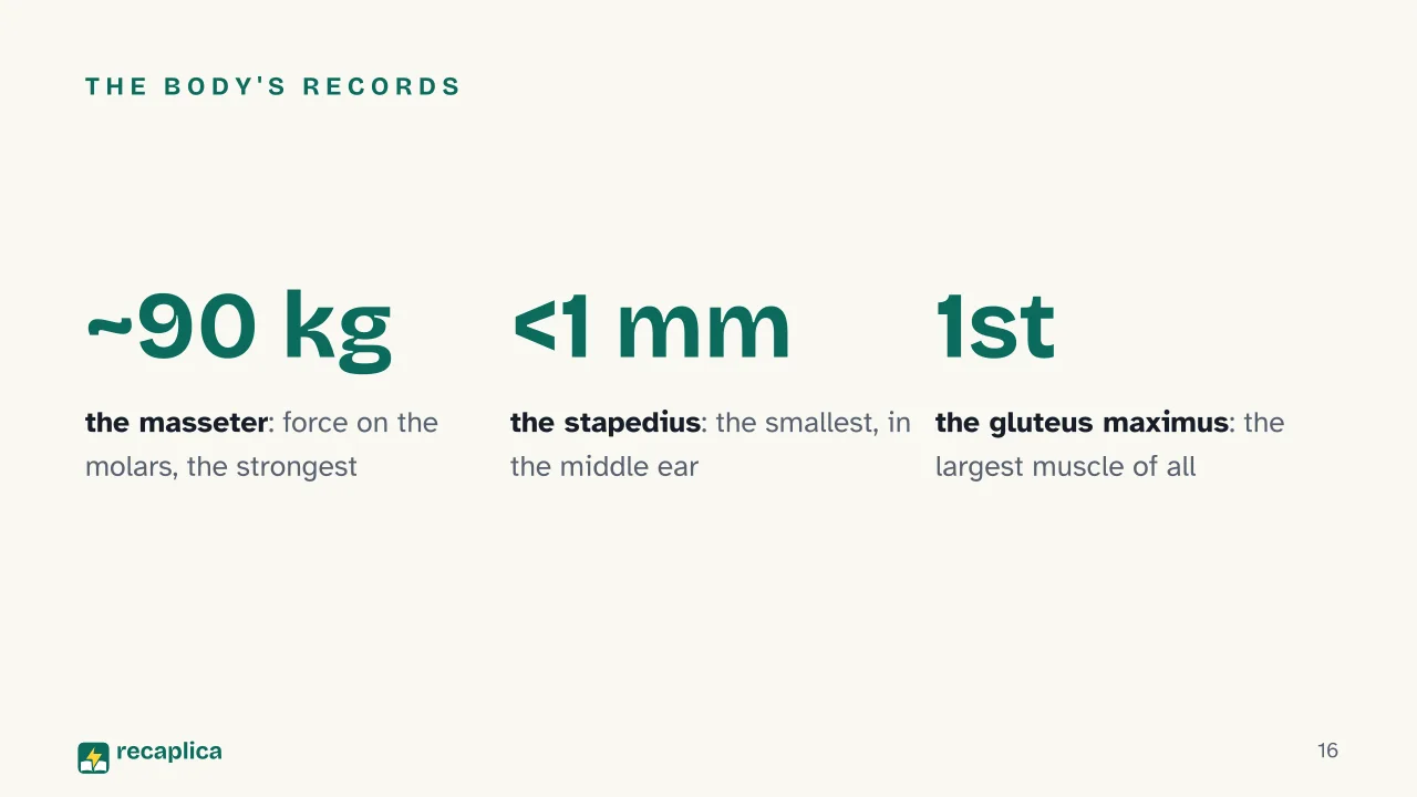
    • ~90 kg the force the masseter, the jaw muscle, can exert on the molars — the highest of any muscle in the body for absolute force Source: Healthline

    Deep Dive

    Three tissues, three different jobs

    Like every other tissue in the body, muscle tissue is made of specialised cells called myocytes. The difference between the three types of muscle comes down mostly to the shape of these cells and how the proteins that generate movement, actin and myosin, are arranged inside them.

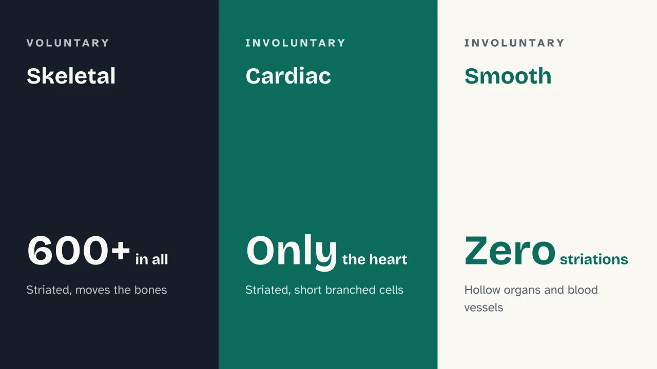
    Skeletal muscle moves the bones and is under voluntary control, even though in some cases it works semi-automatically (think of the diaphragm, which breathes for you even when you’re not thinking about it). Cardiac muscle forms the heart and is involuntary: its cells are short, branched, and linked by junctions called intercalated discs, which spread the electrical signal and make the heart contract in a coordinated way. Smooth muscle, finally, lines the walls of hollow organs such as the stomach, intestines and blood vessels: it is involuntary, and its spindle-shaped cells lack the orderly arrangement of contractile proteins seen in the other two types.

    FeatureSkeletalCardiacSmooth
    Cell shapeLong, cylindrical, unbranchedShort, branchedSpindle-shaped
    NucleiMultiple, at the peripheryOne, centralOne, central
    StriationsYesYesNo
    ControlVoluntaryInvoluntaryInvoluntary
    LocationAttached to bones via tendonsOnly in the heartWalls of hollow organs and blood vessels

    Skeletal muscle: a structure like nested boxes

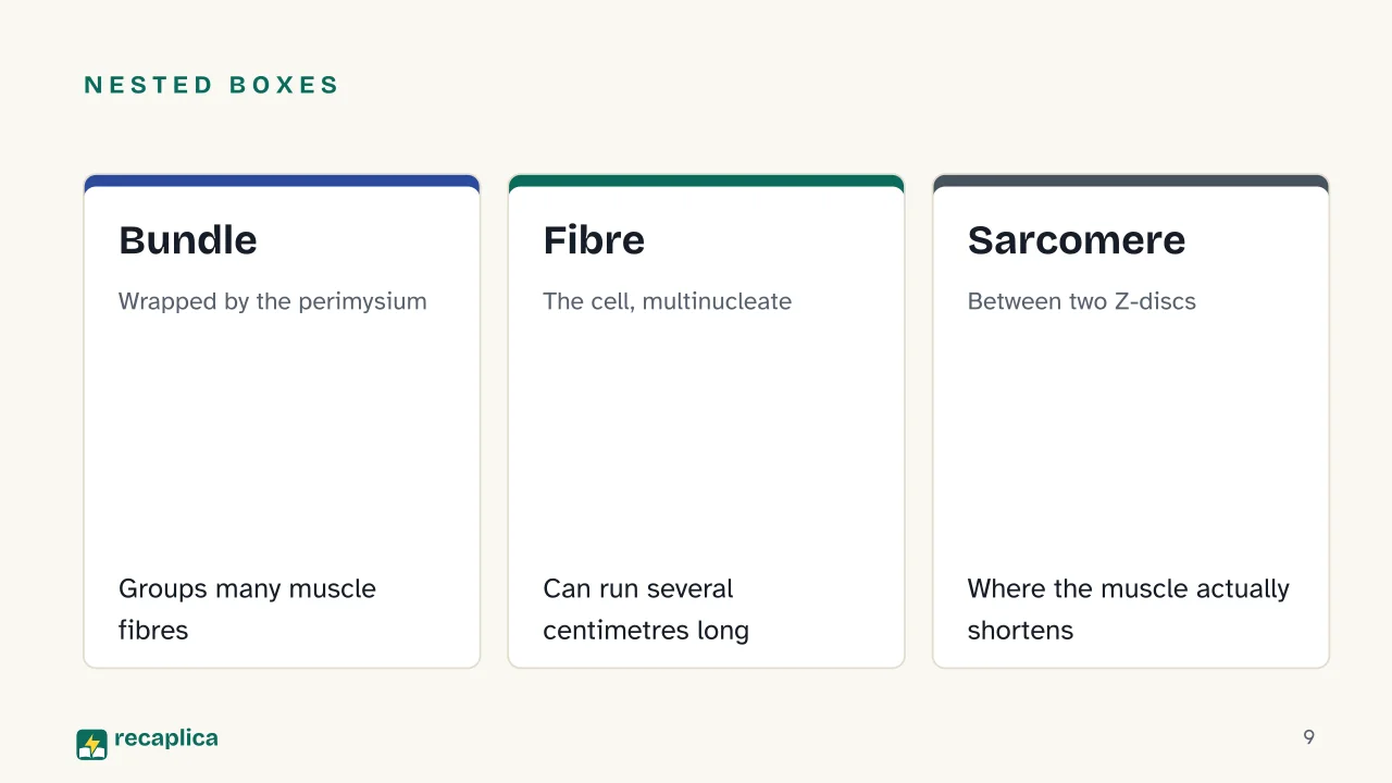
    A skeletal muscle is not a single block: it is a set of layers nested one inside the other. On the outside, a layer of connective tissue called the epimysium wraps the entire muscle. Inside, the muscle is divided into bundles, each wrapped by the perimysium; each bundle contains many muscle fibres (the individual cells), each in turn wrapped by the endomysium.

    The muscle fibre is an unusual cell: it is multinucleate, meaning it contains several copies of DNA inside a single cell that can run several centimetres long. Inside the fibre sit the myofibrils, extremely thin filaments (about a thousandth of a millimetre across) made of sarcomeres lined up one after another.

    The sarcomere, where the muscle actually shortens

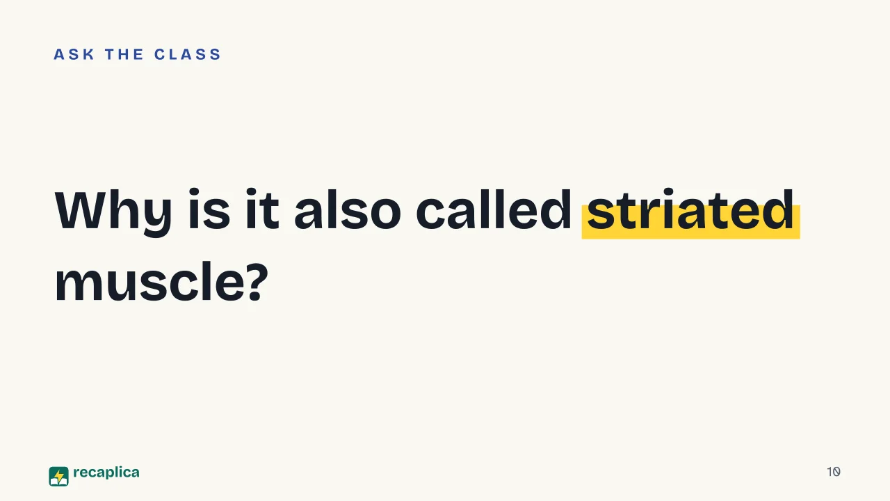
    The sarcomere is the basic contractile unit of skeletal muscle: the stretch of myofibril between two Z-discs. Under a microscope you can see alternating light and dark bands, which is where the name “striated muscle” comes from: the A band, dark, contains the thick myosin filaments, partly overlapped by the thin actin filaments; the I bands, light, sit on either side of the A band and contain only actin; the M line runs through the centre, and near it sits the H zone, where only myosin is present. An elastic filament made of the protein titin holds everything under tension.

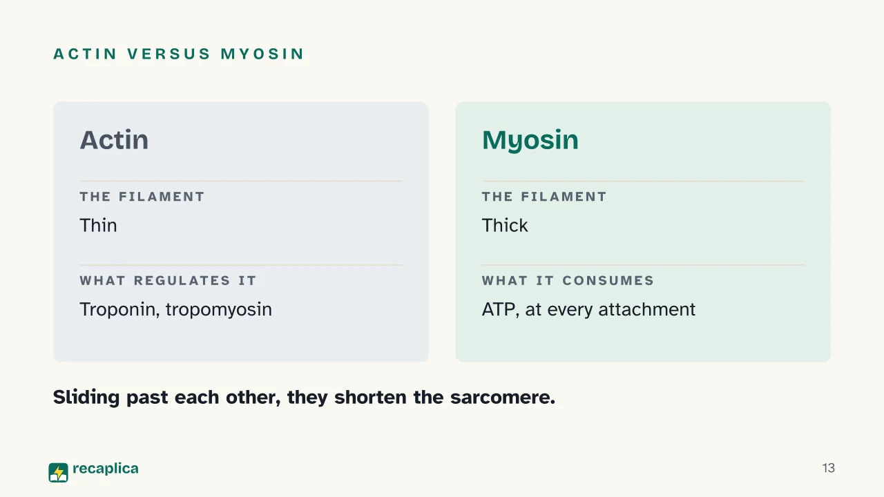
    Two “regulatory” proteins, troponin and tropomyosin, sit on the actin filaments and control when actin and myosin are allowed to link up.

    How a contraction switches on

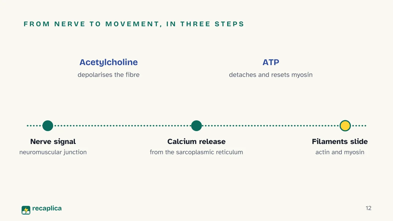
    The signal starts in the nervous system. At the neuromuscular junction, the nerve releases the neurotransmitter acetylcholine, which opens channels on the muscle fibre’s membrane and depolarises it. This electrical signal tells the sarcoplasmic reticulum — a network of internal membranes inside the fibre that stores calcium — to release large amounts of calcium ions.

    Calcium binds to troponin, which in turn shifts tropomyosin and exposes the spots on actin where myosin can attach. From here starts what is known as the cross-bridge cycle: the myosin head attaches to actin, consumes ATP to detach and reset, attaches again a little further along, and pulls the actin filament toward the centre of the sarcomere. Repeated rapidly across thousands of sarcomeres in a row, this sliding shortens the whole fibre: this is the sliding filament theory.

    Practical example: when you pick up a bag of groceries, the signal starts in the brain, travels down a motor neuron, and releases acetylcholine onto the biceps: within a fraction of a second calcium is released, actin and myosin begin sliding past each other, and the arm bends. Stop thinking about it and the nerve signal stops: calcium goes back into the sarcoplasmic reticulum and the muscle relaxes.

    Voluntary or involuntary: who decides

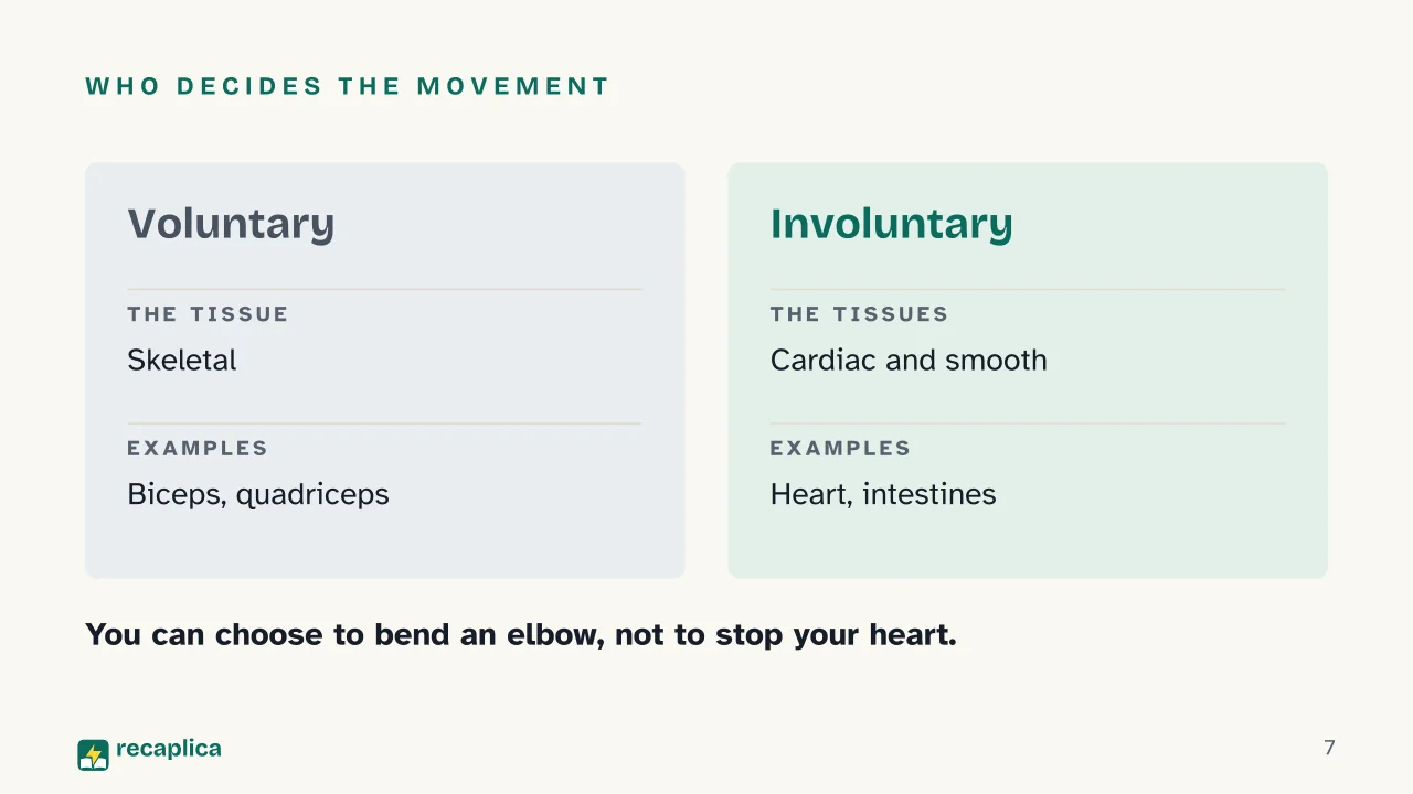
    Only skeletal muscle responds to a conscious command: you can choose to bend an elbow or squeeze an eye shut. Cardiac and smooth muscle work without any need for thought: the heart beats on its own, the intestines push food along, blood vessels narrow or widen to regulate blood pressure, even while you sleep.

    Muscle cells, like all others, still arise from the division of existing cells — the same principle explained in detail for mitosis. In adult skeletal muscle, though, growth happens mostly by enlarging fibres that already exist: that is the mechanism behind muscle hypertrophy, the muscle’s response to training and rest.

    The main muscle groups

    Skeletal muscles are grouped by body region. In the limbs they move the arms and legs — biceps, quadriceps, hamstrings. In the trunk they support posture and protect the organs (abdominals, back muscles); in the head and neck they govern expressions and chewing instead. A special case is the diaphragm, the skeletal muscle that contracts and relaxes automatically to move air in and out of the lungs, while still staying under voluntary control when needed (for example, when holding your breath).

    Among the body’s records: the gluteus maximus is the largest muscle, the masseter (the jaw muscle) is the strongest by absolute force exerted, and the stapedius, in the middle ear, is the smallest of all, measuring less than a millimetre in length.

    Slide deck

    Slides ready to download and make your own in PowerPoint or Google Slides, with speaker notes. Pick the Flash cut or the Full one.
    Slide 1 of the presentation on Muscular system: The muscular systemSlide 2 of the presentation on Muscular system: Why can you raise an arm but not stop your heartbeat?Slide 3 of the presentation on Muscular system: What we will coverSlide 4 of the presentation on Muscular system: Chapter 01: Three tissues, three jobsSlide 5 of the presentation on Muscular system: How much muscle is in a bodySlide 6 of the presentation on Muscular system: Skeletal · Cardiac · SmoothSlide 7 of the presentation on Muscular system: Who decides the movementSlide 8 of the presentation on Muscular system: Chapter 02: Inside skeletal muscleSlide 9 of the presentation on Muscular system: Nested boxes: Bundle, Fibre, SarcomereSlide 10 of the presentation on Muscular system: Why is it also called striated muscle?Slide 11 of the presentation on Muscular system: Chapter 03: How a contraction switches onSlide 12 of the presentation on Muscular system: From nerve to movement, in three stepsSlide 13 of the presentation on Muscular system: Actin versus myosinSlide 14 of the presentation on Muscular system: Chapter 04: False myths and recordsSlide 15 of the presentation on Muscular system: The heart is not made of the stomach's muscle tissue.Slide 16 of the presentation on Muscular system: The body's recordsSlide 17 of the presentation on Muscular system: What is the basic contractile unit of skeletal muscle?Slide 18 of the presentation on Muscular system: And now, the review
    Flash10 slidesThe essential thread, to present in classFull18 slidesEvery chapter and the deeper detail

    Common myths

    • ✗ Myth All muscles move when we decide to move them.

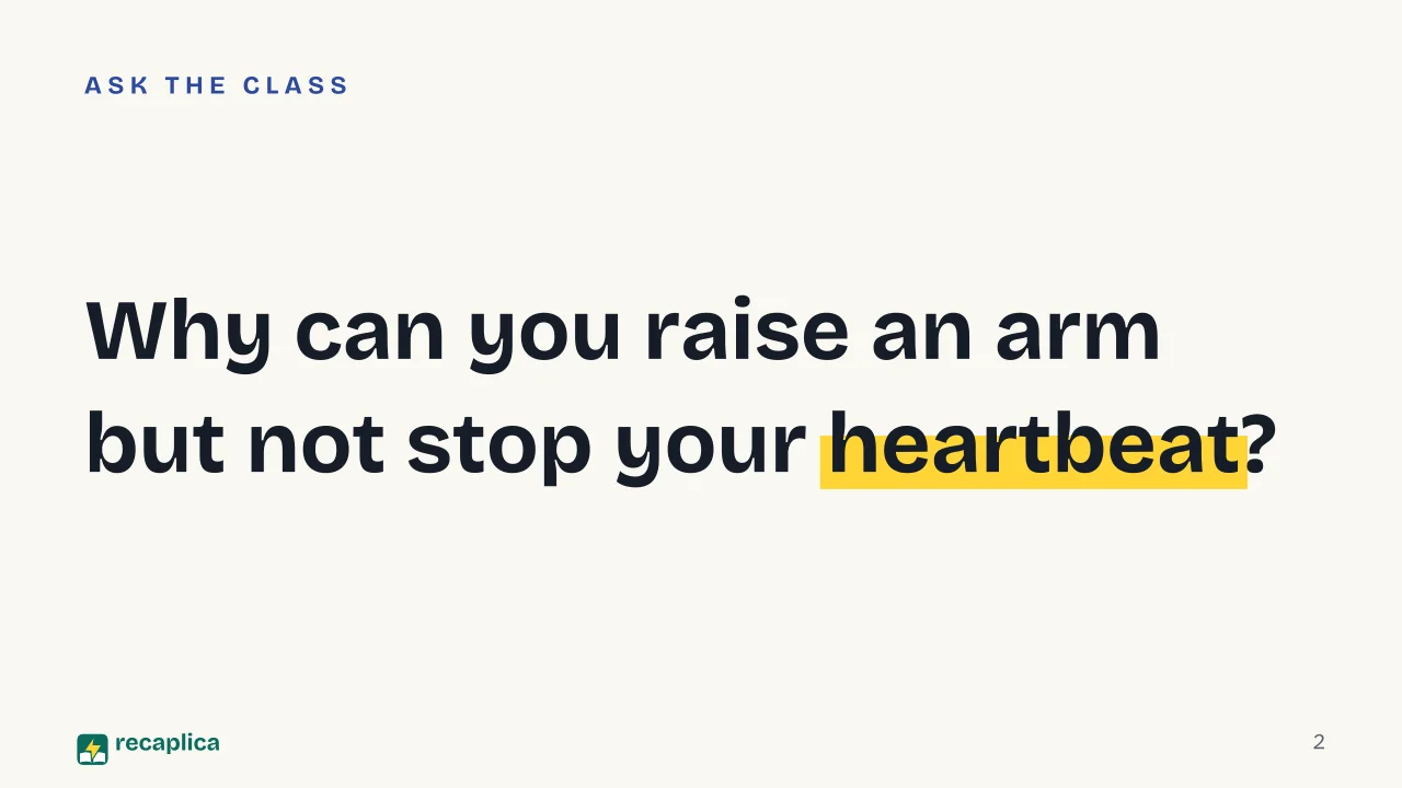
      ✓ Reality Only skeletal muscle is under voluntary control. The heart and the smooth muscle in the stomach, intestines and blood vessels work automatically, with no conscious command needed: that is why you can choose to raise an arm but not to stop your heartbeat.

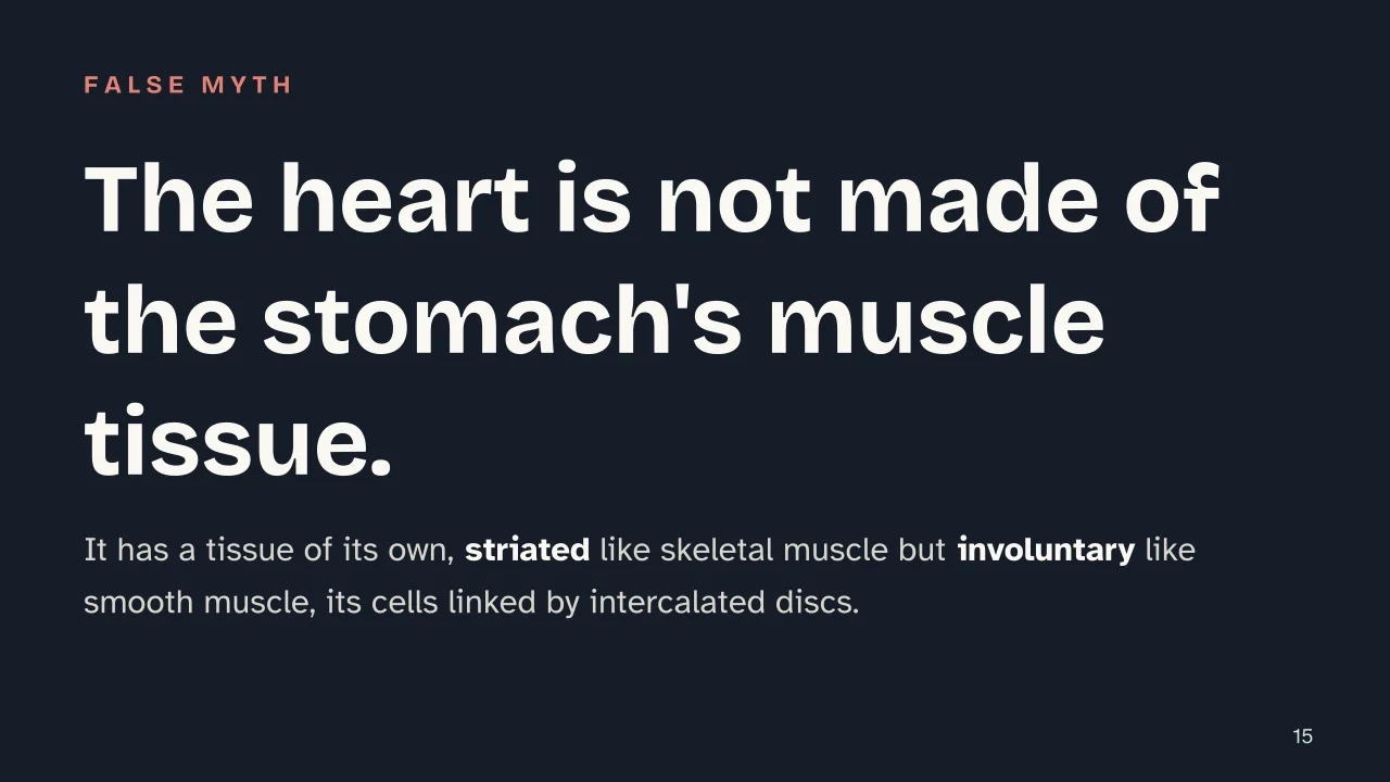
    • ✗ Myth The heart is made of the same muscle tissue as the stomach.

      ✓ Reality The heart has its own tissue type, cardiac muscle: striated like skeletal muscle, but involuntary like smooth muscle. Its cells are linked by junctions called intercalated discs, which spread the electrical signal and make the heart contract in a coordinated way.

    • ✗ Myth The number of muscles in the human body is a fixed figure, the same in every source.

      ✓ Reality The most commonly cited estimate is 'more than 600' skeletal muscles, but the exact number depends on how muscles are counted and grouped: some classifications list over 650. It is not an error, it is a matter of anatomical convention.

    Mind map

    Drag the background to move around and the nodes to reposition them; use − and + to collapse and expand branches.
    Customize
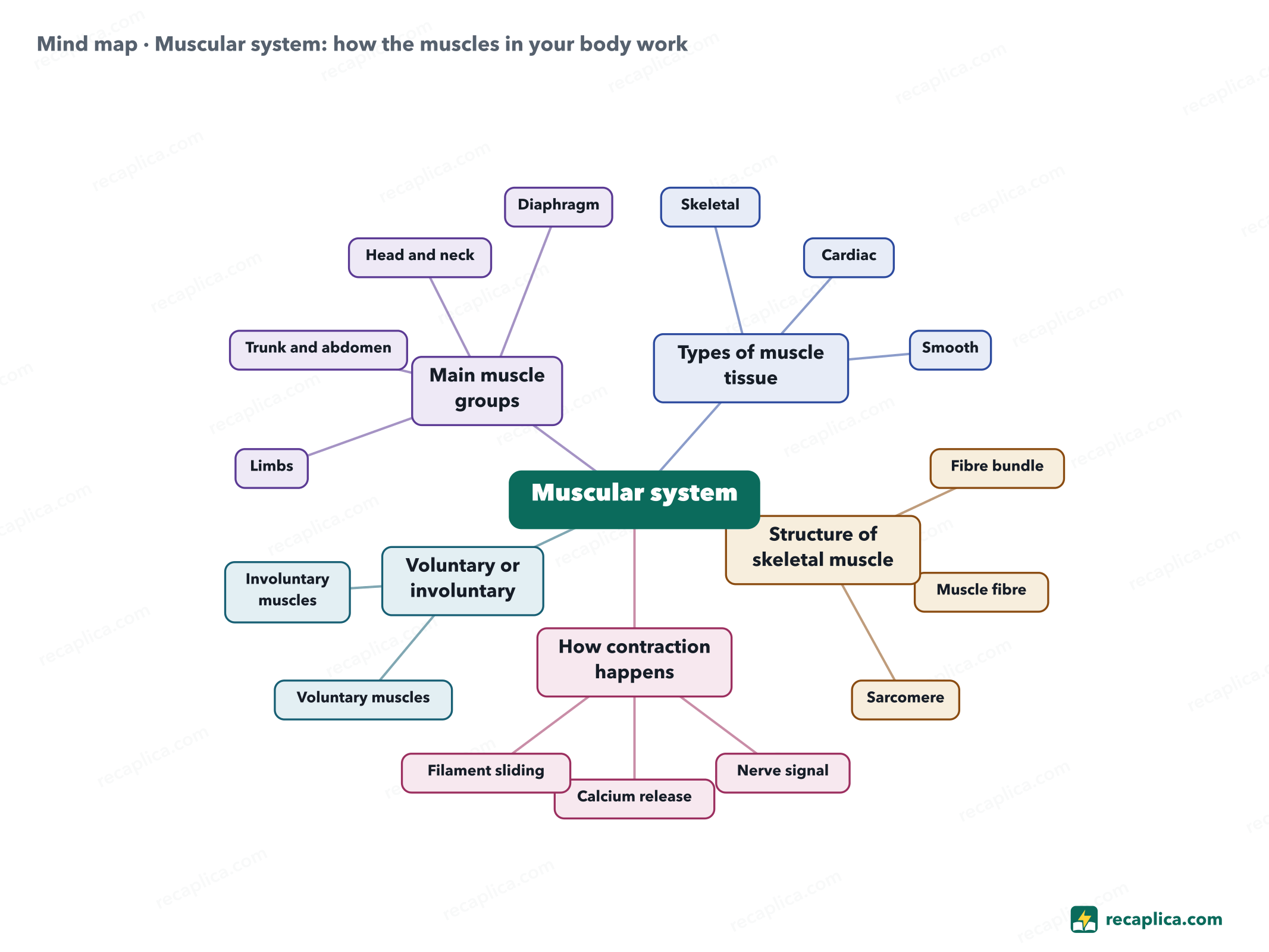
    Mind map: Muscular system: how the muscles in your body work
    • Muscular system
      • Types of muscle tissue
        • Skeletal Striated, voluntary, moves the bones
        • Cardiac Striated, involuntary, only in the heart
        • Smooth Not striated, involuntary, in hollow organs
      • Structure of skeletal muscle
        • Fibre bundle Wrapped by the perimysium
        • Muscle fibre Multinucleate cell, contains the myofibrils
        • Sarcomere Contains the actin and myosin filaments
      • How contraction happens
        • Nerve signal Acetylcholine, depolarises the membrane
        • Calcium release Binds to troponin
        • Filament sliding Actin and myosin, powered by ATP
      • Voluntary or involuntary
        • Voluntary muscles Biceps, quadriceps
        • Involuntary muscles Heart, intestines
      • Main muscle groups
        • Limbs
        • Trunk and abdomen
        • Head and neck
        • Diaphragm

    Quiz: test yourself

    Answer the questions to check what you have learned: you get instant feedback and a short explanation.

    Grade 0/10 0/5
    1 What are the three types of muscle tissue in the human body?

    These are the three recognised muscle tissues: skeletal moves the bones and is voluntary, cardiac forms the heart, smooth lines hollow organs and blood vessels. Cardiac and smooth are both involuntary.

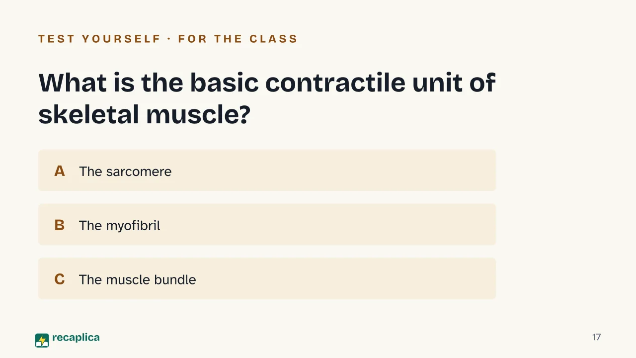
    2 What is the basic contractile unit of skeletal muscle?

    The sarcomere is the stretch of myofibril between two Z-discs: it contains the actin and myosin filaments that shorten it as they slide past each other. The myofibril is the structure made of many sarcomeres in a row, the bundle groups several fibres, and the tendon connects the muscle to the bone.

    3 What triggers the release of calcium inside a muscle fibre during contraction?

    The nerve releases acetylcholine, which depolarises the fibre's membrane; this signal tells the sarcoplasmic reticulum to release its stored calcium, the first step toward actin and myosin linking up.

    4 True or false: cardiac muscle is under voluntary control, like the biceps.

    False: the heart beats on its own, without us deciding when it contracts. It is striated like skeletal muscle, but its control is involuntary, like smooth muscle.

    5 Which muscle is the strongest in the human body by absolute force exerted?

    The masseter can close the jaw with a force of up to roughly 90 kg on the molars. The gluteus maximus is the largest muscle by size, not the strongest in this sense.

    Answers: 1-A · 2-A · 3-A · 4-B · 5-A

    Flashcards

    Tap the card to flip it and check whether you remember the answer, then move to the next one.

    1 / 8

    Explain it in your own words

    The ultimate test: if you can explain it in simple words, you've truly understood it. Write your explanation, then compare it with the Recap.

    Your explanation is saved only on this device.

    The muscular system is the set of muscles that lets you move, breathe and keep your heart beating: the human body has more than 600 of them, and together they make up roughly 30-40% of total body mass. There are three types of muscle tissue — skeletal, cardiac and smooth — each with a different cell structure and a different degree of conscious control. Skeletal muscle is organised into progressively smaller units down to the sarcomere, where two proteins, actin and myosin, slide past each other and shorten the fibre. Contraction starts with a nerve signal, moves through a release of calcium, and runs on energy from ATP.

    Frequently asked questions

    How many muscles are in the human body?

    More than 600 skeletal muscles, according to Cleveland Clinic. The exact number varies depending on how muscles are counted and grouped: Healthline reports over 650 named skeletal muscles, but there is no single, universal count.

    What is the difference between voluntary and involuntary muscle?

    Voluntary muscle, meaning skeletal muscle, contracts when we decide to move it (walking, grabbing an object). Involuntary muscles, cardiac and smooth, work automatically: they keep the heart beating, move food through the intestines, and regulate the width of blood vessels, all without conscious control.

    How does a muscle contract?

    A nerve signal releases acetylcholine onto the muscle fibre, which depolarises and triggers the release of calcium from the sarcoplasmic reticulum. Calcium lets myosin filaments latch onto actin filaments; by consuming ATP, myosin pulls the actin toward the centre of the sarcomere and the muscle shortens.

    Why is skeletal muscle also called striated muscle?

    Because under a microscope it shows alternating light and dark bands, caused by the orderly arrangement of actin and myosin filaments inside the sarcomeres. Smooth muscle lacks this regular arrangement, so it does not appear striated.

    What is the smallest muscle in the human body?

    The stapedius, in the middle ear: it measures less than a millimetre in length. It controls the vibration of the stapes, the smallest bone in the body, and protects hearing by dampening loud sounds.

    Sources

    • Kenhub — Muscles and muscle tissue: Types and functions
    • Kenhub — Sarcomere: anatomy, structure and function
    • Cleveland Clinic — How Many Muscles Are in the Human Body?
    • Cleveland Clinic — Skeletal Muscle (Striated Muscle): What It Is & Function
    • Treccani — Tessuto muscolare (Enciclopedia Treccani)
    • NCBI Bookshelf (StatPearls) — Physiology, Muscle Contraction
    • Healthline — What's the Largest Muscle in the Body?
    • Healthline — How Many Muscles Are in the Human Body?

    Every Recap goes through an independent review before publication.

    Every evening, the day's new Recaps on our Telegram channel. Join the channel →

    Keep learning

    • Science Digestive System: How It Breaks Down Food, Step by Step The digestive system breaks food down, piece by piece, into molecules the body can absorb. It starts in the mouth, where teeth cut food into fragments and saliva carries the first enzymes, continues in the stomach, which churns food with gastric acid, and does most of its chemical work in the small intestine, according to StatPearls (2022). Along the way, the liver, pancreas, and gallbladder — organs outside the digestive tract itself — add bile and enzymes to the mix. What's left moves on to the large intestine, which reclaims water and minerals before waste leaves the body. Read the Recap →
    • Science How the Immune System Works: Barriers, Cells, and Memory The immune system isn't a single organ — it's a network of barriers, cells and organs scattered across the whole body. Skin and mucous membranes form the outer wall, stopping most invaders before they get anywhere. Anything that gets past this wall runs into innate immunity, fast but generic, and then adaptive immunity, slower but aimed at that one invader specifically. Adaptive immunity also leaves something behind: memory, which is why a second encounter with the same pathogen gets a much quicker response. Read the Recap →
    • Science Indigestion Remedies: Symptoms, Causes, and What Really Helps Indigestion, which doctors call dyspepsia, isn't one disease but a group of symptoms that show up together after meals, according to the NIDDK (National Institutes of Health): pain or burning in the upper abdomen, feeling full too soon, bloating. The causes fall into three families, per the same source: a misfiring gut-brain conversation, certain common medications, and organic or infectious stomach problems. Heartburn from acid reflux is a different condition, with a different mechanism. Remedies range from everyday habits to over-the-counter medicine, with a doctor's visit when specific warning signs show up. Read the Recap →

    recaplica

    Clear in 30 seconds, yours in 10 minutes.

    Recaps Mind maps Request a Recap Telegram channel Mind map maker Our method About Privacy & cookies Legal notes & terms of use

    © 2026 Recaplica · A project by Curi S.r.l. — VAT IT05472000750

    Statistics, only if you say so

    To learn which Recaps help most we would use Google Analytics, with aggregate, anonymous data. It starts only with your OK, and you can change your mind anytime. Privacy policy